This new method could allow scientists to gain important insights into what goes wrong when DNA becomes damaged, The Optical Society reported on Friday.
The research, published in the peer-review journal Optica, was led by W. E. Moerner of Stanford University, who carried out the first optical detection and spectroscopy of a single molecule in 1989.
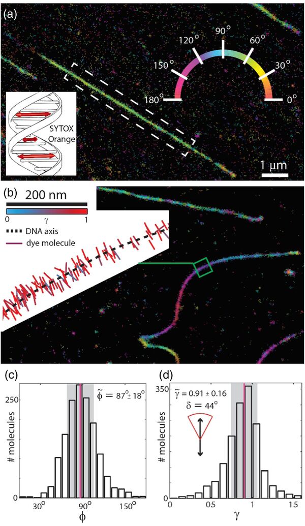
The team has improved this technique by adapting the laser used to analyze thousands of fluorescent dye molecules, which can slide into the areas between the DNA base pairs, attach to the side of the DNA, or connect to it via a floppy tether.
By augmenting this conventional fluorescence microscope with an electro-optic modulator (EOM), they were able to observe the orientation and movement of the dye molecules.

"You can think of these new measurements as providing little double-headed arrows that show the orientation of the molecules attached along the DNA strand," Moerner said.
"This orientation information reports on the local structure of the DNA bases because they constrain the molecule. If we didn't have this orientation information, the image would just be a spot."
"If someone has a single-molecule microscope, they can perform our technique pretty easily by adding the electro-optic modulator," said Adam Backer, lead author of the paper.
"We've used fairly standard tools in a slightly different way and analyzed the data in a new way to gain additional biological and physical insight."


